- Understand PCI-AI’s three-tier p53 expression framework: null, wild-type, and diffuse
- Recognize how each expression pattern maps to the underlying mutation type
- Learn how TriControl™ and DualControl™ panels provide reference standards for each tier
What is p53?
p53 is a protein commonly referred to as the “Guardian of the Genome,” whose main function is to protect the integrity of DNA within a cell. p53 is made from a gene called TP53The gene that codes for the p53 protein, one of the most frequently mutated genes across human cancers., and is classified as a tumor suppressor, as its normal function is to stop tumors from forming.
Mechanically speaking, p53 acts as a transcription factor, meaning it turns other genes on or off in response to various factors, such as cellular stress, DNA repair, or apoptosis. p53 is like the quality control inspector of a genome: when a cell’s DNA gets damaged, p53 assesses the damage and will either pause the cell to allow for repairs, or trigger the cell to self-destruct rather than risk becoming cancerous.
TP53 is the most frequently mutated gene found across human tumors. For example, a rare, heritable condition called Li-Fraumeni syndrome develops when an individual only inherits one functional copy of the p53 gene, predisposing them to developing tumors across different tissues in early adulthood.
What is Immunohistochemistry (IHC)?
IHC is a lab technique that utilizes antibodies to detect specific proteins, or antigens, in a tissue sample. Similarly to how the immune system makes antibodies to find and destroy harmful substances, IHC uses that antigen-finding ability to diagnostically detect proteins in a tissue sample instead.
Mechanically, an antibody that only binds to specific target proteins (p53, for example) is applied to a sample. This is followed by a chemical reaction that makes that binding visible under a microscope. IHC preserves the surrounding tissue architecture while showing exactly where a target protein is located — including whether it appears as nuclear expressionProtein staining localized to the cell nucleus, as opposed to the cytoplasm or cell membrane. — making it incredibly valuable to pathologists.
p53 Applied
p53 is one of the most studied biomarkers in pathology, and its IHC staining pattern is read using a three-tier classification that closely tracks the underlying gene’s mutation status. Because p53 is mutated in such a wide range of cancers, this pattern has become a very practical alternative to genetic testing. There are three different patterns of p53 that are clinically tested for: wild-type, null, and overexpressed (diffuse) patterns.
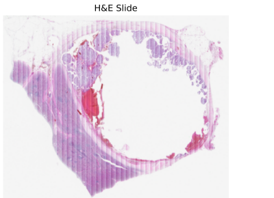
Wild-type (normal) — shows up at a low, variable intensity, typically present in less than half of tumor cells. This is the pattern expected when TP53 is not mutated, and the protein is continuing its normal and expected function within the cell.
Null — a complete absence of nuclear p53 staining, despite positive internal positive controlsNormal adjacent tissue used to confirm the staining procedure itself worked, so a “null” result can be trusted as biological rather than a technical failure. confirming the test’s accuracy. This pattern typically points to a truncating mutationA mutation that cuts a protein short, often preventing it from being made or functioning at all. — a mutation that stops the cell from producing any functional protein at all.
Diffuse / overexpressed — strong, uniform staining across at least 80% of tumor cells. This also signifies a mutation, but a different kind: certain mutations produce a dysfunctional p53 that accumulates in the cell rather than dissipating, leading to an overabundance of protein.
ⓘ Colors here indicate classification category, not actual stain color.
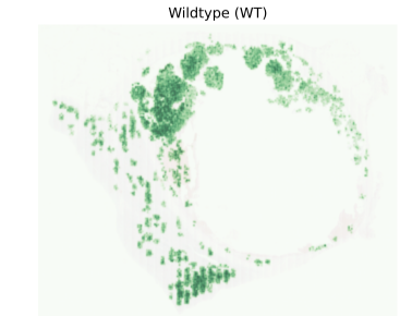
Look for: scattered, low-intensity staining covering less than half the tissue.
TriControl™ covers all three patterns
PCI-AI’s TriControl™ panel includes null, wild-type, and diffuse cell lines — providing a physical reference standard for each p53 expression category described above.
View TriControl™ →Clinical Relevance
Pathologists treat abnormal p53 IHC patterns as a reliable surrogate markerA practical, faster stand-in for a more complex or expensive test — here, p53 IHC pattern standing in for direct TP53 sequencing. for an underlying TP53 mutation. In endometrial cancer specifically, p53 status has been incorporated into the WHO-endorsed molecular classification system.
In the WHO molecular classification of endometrial cancer, p53-abnormal tumors represent a subgroup of patients with the poorest prognosis and the greatest likelihood of benefiting from additional treatment.